Did you know that live worms can be used to heal wounds/injuries?

Some part of our body, such as the skin or bones can grow new tissue to repair damage, but nerve cells are not easily replaced. They have to repair themselves, but this process is inefficient and slow, if it happens at all. This poses a major hurdle for recovery after injuries to the nervous system, such as traumatic brain injury, spinal cord injury, eye injury, neurodegenerative disease or stroke.
Nerve cells, or neurons, are the basic unit of the nervous system, and billions of them form intricate circuits that are essential for our body’s day-to-day functions. They are unlike any other cells in our body because of their unique cellular structure. A neuron has a long, thin, cable-like structure known as the axon, which transmit electrical messages to target tissues, like muscles. When axons are injured, they can no longer transmit messages and function is affected. The neuron needs to repair that connection to re-establish the line of communication.
To restore function when a neuron’s axon is severed in two, the section attached to the main cell body known as the proximal fragment needs to successfully regrow and reconnect with its original target tissue. The easiest option would be to reconnect to the distal fragments that is, the part of the axon separated from the main cell body. But normally after injury, in humans and other mammals, this distal fragment withers and dies, and the proximal axon is left with the difficult, if not impossible, task of regrowing a full length to reach its original target tissue.
However, in many invertebrates such as the sea slug, earthworm, leech, crayfish and the roundworm (C. elegans), the easiest option is reconnecting the two broken halves of the axon, a repair process known as axonal fusion allows the two fragments of the severed axon to fuse back together, preventing degeneration of the distal fragment and rapidly restoring the neuron’s function. This is much more efficient than regrowing the axon all the way back to original target tissue. So, there is enormous potential for us to learn how these animals spontaneously repair their axons and apply our knowledge to promote nerve repair in humans.
The studies of animal models, including roundworms(C. elegans), flies, zebrafish and rodents, which can easily be genetically modified, allow scientists to learn more about the molecules involved in many biological processes, including healing wounds or neuron repair, and how they affect different cells and tissue types within a whole organism.
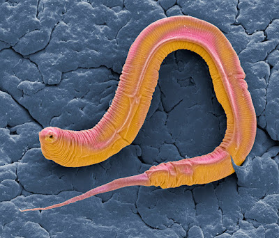
Sydney Brenner, introduced roundworm as an experimental animal model to study development and neurobiology. Roundworm (C. elegans) has only 959 cells and exactly 302 neurons, so its simple biology makes it easier for us to identify the molecules involved in biological processes. At a genetic level, roundworm (C. elegans) shares about 60-80% of the same genes as humans, which means that similar molecules involved in neuron repair are likely to be found in humans.
A study on the molecular mechanisms involved in axonal fusion in Roundworm(C. elegans), published in Nature in 2015. Shows that a neuron responds to injury by changing the composition of the lipid component of its cell membrane, which acts as a ‘save me’ signal, the presence of surface wounds triggers another series of chemical reactions that allow the worms to quickly close cuts in their surfaces that would turn fatal if left unrepaired. The severed neuron is then repaired by axonal fusion through a protein known as EFF-1.
"Andrew Chisholm, a professor of biology at UC San Diego, said a biochemical pathway that involves calcium is activity wound in the worms, “It’s been known for some time that one of the things that happens when you damage a cell is that calcium levels within the cell increase.”
But in a series of experiments with C. elegans, Chisholm and postdoctoral fellow Suhong Xu found out much more. They took time-lapse movies of areas around the transparent worms where they punctured the skin with a needle or laser. Then they monitored the calcium with a fluorescent protein so they could see how the calcium molecules spread from the point of injury. They also developed genetic screens to pinpoint the specific calcium pathway or “channel” that is signaling the presence of the wound and stimulating the healing process.
“We think the channel is playing an important role in either sensing damage or responding to some other receptor that senses damage,” said Chisholm. “Is it sensing a change in the tension of the cell? Or Is it sensing some kind of change in electrical potential? We don’t know.”
“They have a hydrostatic skeleton in which the skin and muscles are under pressure to allow the animal to stay semi-rigid, so when you jab a worm with a needle it will, in effect, explode,” he said. “But remarkably, they don’t die when you do that because they have evolved ways to very rapidly close wounds to survive in the wild. In their natural environment, their predators try to exploit the worm’s vulnerable exoskeleton. There are a whole group of fungi with tiny spikes that just sit around waiting for the worms to crawl over them so they can poke holes through their cuticle.”
“We think that calcium is regulating this process,” said Chisholm, “because if you knock out calcium with a drug that chelates calcium, essentially locking it up, you don’t get the ring. If you have a genetic mutant worm with low levels of calcium, you don’t get the ring. But if you bathe this mutant in calcium, you can restore this ring.”
In addition, the researchers discovered in roundworms that a protein called DAPK-1 acts to inhibit the closure of wounds, raising the possibility that drugs that inhibit the action of this protein could improve the wound healing process in humans.


Comments
Post a Comment